The connection between hyperbaric oxygen therapy and burns is not mysterious. It comes from a single observation made in the 1950s about what happens at the edges of a burn — and from the awkward fact that the tissue there does not stay the same size. It moves. Sometimes it shrinks. Sometimes it expands over the following days.
That expansion is the whole story.
A note on scope before anything else: this article is educational. Thermal injury is managed in hospital settings by qualified professionals using hospital-grade equipment and hospital protocols. Non-medical hyperbaric chambers — the mild, low-pressure kind used for general wellness, recovery, and lifestyle purposes — are not intended for burn injury and are not a substitute for professional care. Fire-related exposures other than the thermal injury itself follow an entirely separate logic and fall outside this discussion. Nothing here is guidance for handling a burn.
Step One: Heat Does Not Stop When the Heat Source Leaves
A thermal burn starts as direct thermal damage. Proteins denature. Cell membranes fail. That part is immediate and mechanical, and it’s the part everyone pictures.
What happens next is less obvious. Heat also injures the small vessels feeding the surrounding tissue — capillaries, venules, the fine plumbing that keeps a few cubic millimeters of skin alive. Those vessels leak. Endothelial cells swell, gaps open between them, and plasma moves into the interstitial space [1].
Fluid leaving the vessels does two things at once:
- Edema builds. Pressure rises inside the tissue compartment.
- Perfusion drops. Less volume moving through narrowed, sludgy capillaries.
Now you have a supply problem. Oxygen delivered by blood is the only oxygen that tissue receives, and blood flow just fell while diffusion distance just increased. The result is regional tissue hypoxia — not everywhere, but specifically in the band of tissue surrounding the destroyed core.
Hypoxia triggers inflammation. Neutrophils arrive, adhere to damaged endothelium, and release reactive species and proteases as part of a normal response to injury. Useful in principle. In a tightly packed, already-starved margin of tissue, that response adds a second layer of stress on cells that were surviving the first one [2].
Heat damage → vessel damage → swelling and low flow → low oxygen → inflammation. Each step feeds the next.
Step Two: The Zone of Stasis
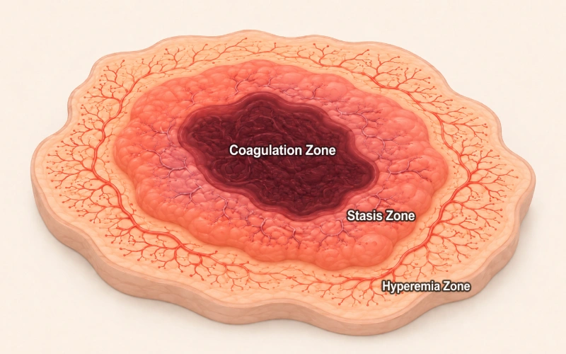
In 1953, a surgeon described burn wounds as three concentric zones rather than one uniform area [3]. The model has held up remarkably well and it still frames how researchers think about the problem.
| Zone | What it is | Oxygen status | Fate |
|---|---|---|---|
| Coagulation | Central area of direct thermal destruction | None — tissue is non-viable | Irreversible from the outset |
| Stasis | Surrounding band with damaged microvasculature, sluggish flow, marked edema | Hypoxic; delivery is compromised but not absent | Uncertain — may recover, may progress to necrosis |
| Hyperemia | Outer margin with increased blood flow | Well oxygenated | Usually recovers |
The middle row is where every research question lives.
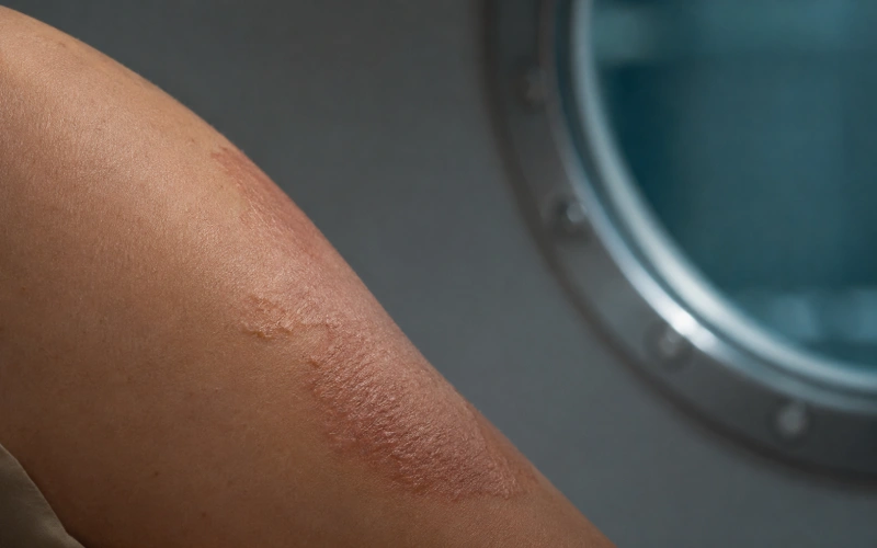
Tissue in the zone of stasis is injured but not dead. Over roughly the first 24 to 72 hours, some of it recovers perfusion and survives. Some of it does not, and the wound quietly gets deeper and wider — a process usually described as burn wound progression or conversion [2]. A wound that looked partial-thickness on day one can look considerably worse on day three without anything new happening to it.
So the question becomes narrow and specific: is stasis-zone tissue salvageable, and can anything shift the odds during that window?
Step Three: Why Hyperbaric Oxygen Entered the Conversation
Once you frame the problem as “hypoxic, swollen, inflamed tissue with a short decision window,” hyperbaric oxygen becomes an obvious thing to test. Not because it’s exotic. Because the proposed mechanisms line up with the specific failures listed above.
Four of them get cited most often.
1. Oxygen availability independent of hemoglobin
Under normal conditions, hemoglobin carries almost all oxygen and it’s already close to saturated. You cannot load much more onto it. What you can do is dissolve oxygen directly in plasma, and that quantity follows pressure in a straightforward, linear way [4].
The numbers make the argument better than the sentence does:
| Breathing condition | Dissolved oxygen in plasma (approximate) |
|---|---|
| Air, 1 ATA | ~0.3 mL/dL |
| High-concentration oxygen, 1 ATA | ~1.5 mL/dL |
| High-concentration oxygen, 2 ATA | ~4 mL/dL |
| High-concentration oxygen, 3 ATA | ~6 mL/dL |
That last row matters because resting tissue oxygen extraction sits in roughly the same range — around 5 to 6 mL/dL. In other words, the dissolved fraction alone becomes physiologically meaningful only once pressure is applied. That is the entire reason pressure appears in this discussion at all, rather than oxygen concentration by itself.
Higher dissolved oxygen also extends the distance oxygen diffuses from a functioning capillary into surrounding tissue [4]. In a region where capillaries are sparse, compressed, or intermittently blocked, diffusion distance matters more than total flow.
2. Reduced edema
This one seems backwards at first. High partial pressures of oxygen cause vasoconstriction — vessels narrow. Narrowing reduces the volume of fluid pushed into leaky tissue, which lowers interstitial pressure. Because the oxygen content of what still arrives is much higher, the reasoning is that delivery is maintained despite reduced flow [4].
Less swelling also means less mechanical compression of the very capillaries the stasis zone depends on.
3. Microcirculatory support
Improved oxygenation of endothelial cells, reduced red cell aggregation, and better preservation of capillary patency have all been described in laboratory and animal work on ischemic tissue. The intent is to keep marginal capillaries open through the window when they would otherwise shut down permanently.
4. Inflammation modulation
Hyperoxia has been shown to interfere with neutrophil adhesion to injured endothelium — a specific, measurable step in the inflammatory cascade [4]. Fewer adherent neutrophils in a hypoxic margin plausibly means less collateral damage to cells that were going to make it.
Putting it together
| Proposed mechanism | Target problem in the burn wound | Where the evidence mostly comes from |
|---|---|---|
| Elevated dissolved plasma oxygen, longer diffusion distance | Tissue hypoxia in the stasis zone | Physiology and animal models; well established as a physical effect |
| Hyperoxic vasoconstriction | Edema and interstitial pressure | Animal models; one small human volunteer study reported reduced wound exudate and swelling [5] |
| Capillary preservation | Reduced perfusion | Animal and laboratory work |
| Reduced neutrophil adhesion | Secondary inflammatory injury | Laboratory and animal work [4] |
| Fibroblast activity, collagen cross-linking, new vessel formation | Later repair phase | Wound biology literature; oxygen is a rate-limiting substrate [6] |
If those four effects hold in a real burn wound, the expected consequence is less secondary tissue injury — a stasis zone that converts to necrosis less often, and a wound that stays closer to its day-one appearance.
Step Four: The Repair Phase
Suppose some stasis-zone tissue is preserved. Closure is also oxygen-dependent. Collagen cross-linking requires oxygen for proline and lysine hydroxylation, fibroblast proliferation is oxygen-sensitive, new vessel formation responds to oxygen gradients, and the oxidative burst immune cells use against bacteria consumes it directly [6].
This part of the argument is more settled than the acute-phase part. Oxygen is a substrate, and wounds in poorly oxygenated tissue behave differently from wounds in well-oxygenated tissue across many wound types.
Step Five: What the Documented Outcomes Actually Show
Here’s where the tidy chain of reasoning meets messy data.
Animal studies have been broadly favorable — reduced edema, better preserved dermal perfusion, less wound progression. Consistent enough that the mechanistic story stayed alive for decades.
Human data are thinner and less unified. The trials that exist are small, run at different eras and different centers, with different pressures, session frequencies, timing after injury, and outcome definitions. Some reported shorter healing times or reduced fluid requirements. Others found no meaningful difference. A systematic review examining randomized human trials in thermal injury concluded that the available evidence was insufficient to support or refute routine use, largely because of trial size and methodological limitations [7].
Work published since has mostly consisted of small series using perfusion imaging endpoints rather than large randomized designs, which has added mechanistic detail without changing that overall verdict.
Which is not the same as “no effect.” It means the studies were not built to detect one reliably.
Meanwhile, hospital-based hyperbaric programs and professional societies have generally kept thermal injury on their lists as an adjunctive consideration, with the reasoning laid out in review literature: the mechanisms are coherent, the animal data support them, the human data are suggestive but incomplete, and decisions are made case by case rather than by rule [8].
Three practical variables come up repeatedly in that literature:
| Variable | Why it constrains use |
|---|---|
| Timing | The stasis zone declares itself within the first 24–72 hours. Anything intended to influence conversion has to fall inside that window, which is logistically demanding to arrange. |
| Logistics | Managing someone with a significant burn inside a pressurized chamber complicates fluid management, airway access, monitoring, and staffing simultaneously. |
| Risk | Pressure-related ear and sinus discomfort, barotrauma, and oxygen-related concerns are real and require trained supervision throughout. |
None of that is trivial, and it explains why the practice is limited to specialized settings rather than widespread.

The Honest Summary
The chain of reasoning is clean:
thermal injury → microvascular damage → edema and reduced perfusion → hypoxia → inflammation → a stasis zone that may or may not survive → oxygen availability as the variable most plausibly under external influence → possible reduction in secondary injury → support for the repair phase
The evidence is not as clean. Promising, mixed, and dependent on protocol details that vary between studies.
The consistent conclusion across the serious literature is the same one worth repeating: where it is used, hyperbaric oxygen is positioned as an adjunct alongside conventional burn management. Not a replacement for it. Fluid resuscitation, wound care, infection control, surgical decisions, and nutrition remain the foundation. Oxygen availability is, at most, one supporting variable.
Where Non-Medical Chambers Fit — and Where They Don’t
This distinction gets blurred constantly online, so it’s worth stating plainly.
| Hospital-grade hyperbaric systems | Non-medical / mild chambers | |
|---|---|---|
| Typical pressure | Roughly 2.0–2.8 ATA | Roughly 1.3–1.5 ATA |
| Oxygen source | Regulated high-concentration delivery systems | Oxygen concentrator, lower concentration |
| Supervision | Trained hyperbaric staff, defined protocols | Owner or facility operator |
| Intended use | Conditions handled in hospital settings | General wellness, recovery, lifestyle, sports and travel-related use |
| Appropriate for burn injury | Decided by qualified professionals | No |
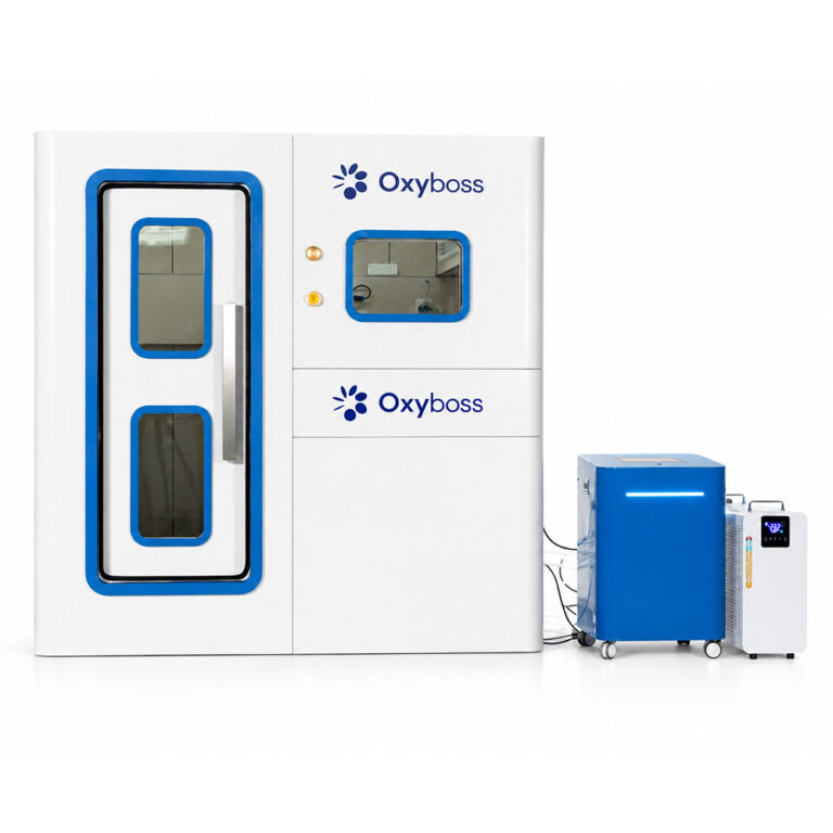
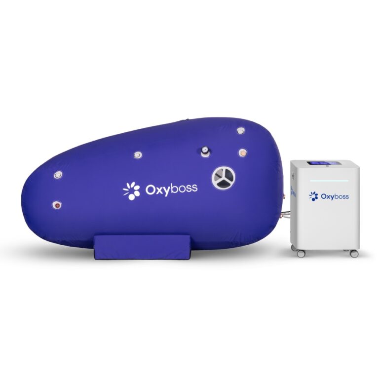
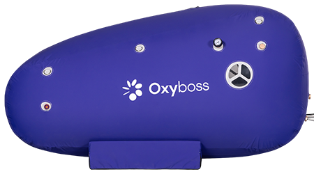
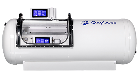
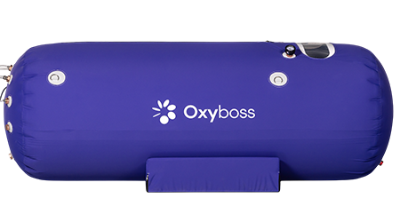
Mild chambers operate at a fraction of the pressure used in the studies discussed above, without the oxygen concentrations, monitoring, or protocol structure those studies involved. Look back at the dissolved-oxygen table: at 1.3 ATA with concentrator-grade oxygen, the plasma fraction stays close to the second row, nowhere near the third or fourth. The research described here does not transfer. Anyone dealing with a burn needs professional care, immediately, in a facility equipped for it.
FAQ
Why is the zone of stasis considered so important? Because it is the only part of a burn wound whose outcome is genuinely undecided. The central destroyed area cannot be recovered, and the outer margin generally recovers on its own. The stasis zone sits between them and can go either way over the first few days — which makes it the only realistic target for anything intended to limit how deep or wide a wound becomes.
If the physiology makes sense, why is the human evidence still mixed? Small trial sizes, mostly. Add wide variation in pressures used, number and spacing of sessions, how soon after injury sessions began, and how “healing” was measured across studies. Aggregating results from studies that differ that much produces weak conclusions even when individual studies look encouraging.
Does breathing high-concentration oxygen at normal pressure achieve the same thing? Not to the same degree. Hemoglobin is already nearly saturated at normal pressure, so the additional oxygen carried in plasma is limited — roughly 1.5 mL/dL against about 6 mL/dL at 3 ATA. The dissolved fraction scales with pressure, and the increase in diffusion distance follows from it.
Could edema reduction be the main mechanism rather than oxygen delivery? Possibly, and some reviewers have argued exactly that. Lower interstitial pressure keeps marginal capillaries open, which improves perfusion, which improves oxygenation — the mechanisms are entangled and hard to separate experimentally. Existing studies were not designed to isolate one from the others.
Is thermal injury an accepted use for hyperbaric oxygen? It appears on the adjunctive lists maintained by hyperbaric professional bodies, meaning it is considered alongside standard management rather than instead of it, and only in appropriately equipped hospital settings. It is not something decided by an individual or attempted at home.
Why would a non-medical site write about this at all? Because the pressures and oxygen concentrations in this research are the clearest available yardstick for equipment boundaries. The 2.0–2.8 ATA range used in hospital studies is not a stronger version of the 1.3–1.5 ATA range found in wellness chambers; they are different application categories with different physics behind them. Explaining the first accurately is what keeps the second from being described in vague language.
References
- Hettiaratchy S, Dziewulski P. ABC of burns: pathophysiology and types of burns. BMJ. 2004;328(7453):1427–1429. https://www.ncbi.nlm.nih.gov/pmc/articles/PMC421790/
- Shupp JW, Nasabzadeh TJ, Rosenthal DS, Jordan MH, Fidler P, Jeng JC. A review of the local pathophysiologic bases of burn wound progression. Journal of Burn Care & Research. 2010;31(6):849–873. https://doi.org/10.1097/BCR.0b013e3181f93571
- Jackson DM. The diagnosis of the depth of burning. British Journal of Surgery. 1953;40(164):588–596. https://pubmed.ncbi.nlm.nih.gov/13059343/
- Thom SR. Hyperbaric oxygen: its mechanisms and efficacy. Plastic and Reconstructive Surgery. 2011;127(Suppl 1):131S–141S. https://www.ncbi.nlm.nih.gov/pmc/articles/PMC3058327/
- Niezgoda JA, Cianci P, Folden BW, Ortega RL, Slade JB, Storrow AB. The effect of hyperbaric oxygen therapy on a burn wound model in human volunteers. Plastic and Reconstructive Surgery. 1997;99(6):1620–1625. https://pubmed.ncbi.nlm.nih.gov/9145133/
- Sen CK. Wound healing essentials: let there be oxygen. Wound Repair and Regeneration. 2009;17(1):1–18. https://www.ncbi.nlm.nih.gov/pmc/articles/PMC2704133/
- Villanueva E, Bennett MH, Wasiak J, Lehm JP. Hyperbaric oxygen therapy for thermal burns. Cochrane Database of Systematic Reviews. 2004;(3):CD004727. https://doi.org/10.1002/14651858.CD004727
- Cianci P, Slade JB, Sato RM, Faulkner J. Adjunctive hyperbaric oxygen therapy in the treatment of thermal burns. Undersea and Hyperbaric Medicine. 2013;40(1):89–108. https://pubmed.ncbi.nlm.nih.gov/23397872/
- U.S. Food and Drug Administration. Hyperbaric Oxygen Therapy: Get the Facts. https://www.fda.gov/consumers/consumer-updates/hyperbaric-oxygen-therapy-get-facts
- Undersea and Hyperbaric Medical Society. Indications for Hyperbaric Oxygen Therapy. https://www.uhms.org/resources/hbo-indications.html